20 016 968

Seeing is living
ReLEx® smile is the new laser technique by Carl Zeiss for the gentle correction of vision defects. It is a minimally invasive treatment method which combines the extensive experience and superior safety of traditional vision correction techniques with numerous innovative benefits, high precision levels and perceptibly greater comfort during the treatment itself.
Our eyes are our most important sensory organ. The human brain obtains over 80 % of its information via the sense of sight. Our eyes are our windows to the world. Seeing is recognizing. Seeing is experiencing. Seeing is independence and freedom. Seeing is living. Well over half the world’s population relies on glasses or contact lenses to see well. But many find that being dependent upon optical appliances interferes with their professional lives and leisure time. Simply being able to see. Without glasses or contact lenses. Completely free of any optical appliances. This is now possible in increasing numbers of cases thanks to ongoing developments in medicine and technology. Refractive visual correction techniques have been scientifically recognized and clinically tested over the last few decades. They have come to represent an important alternative to traditional correction methods such as glasses and contact lenses.

“Perfection is Achieved Not When There Is
Nothing More to Add, But When There Is
Nothing Left to Take Away”
/Antoine de Saint-Exupéry/
The shortest man-made phenomenon
for creating outstanding vision
No microkeratome, no ablation products – these are the inherent advantages of ReLEx® SMILE and ReLEx flex. Because both techniques make exclusive use of the Carl Zeiss VisuMax® femtosecond laser and its superior individual components – giving you access to the benefits of high-precision femtosecond technology: high-level reproducibility and predictability – even in the case of high corrections.
Precise and sensitive treatment VisuMax femtosecond laser
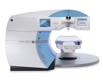
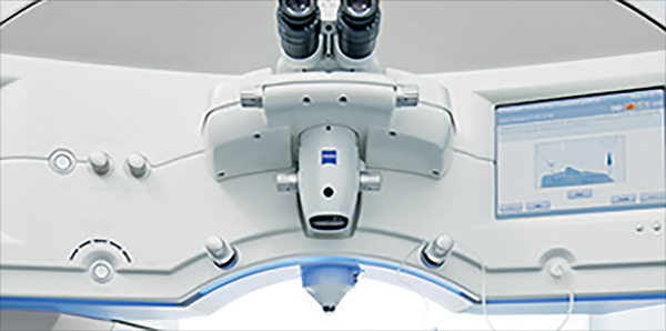
The specially developed laser system used for the ReLEx SMILE procedure is the Carl Zeiss VisuMax® femtosecond laser. In FemtoLASIK surgery it has already impressed patients and physicians alike with its sophisticated technology, its precision and its reliability. ReLEx SMILE using the VisuMax now permits accuracy down to nearest micrometer precision in the preparation of the lenticule within the intact cornea.
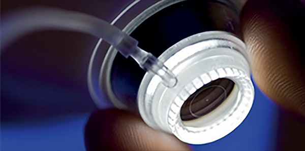
The high-precision femtosecond laser provides targeted correction of the vision defect while leaving the surrounding corneal tissue virtually unaffected. A contact glass specially designed to match the individual anatomy of the cornea permits customized treatment in which the corneal tissue does not need to be unnecessarily compressed.
An ergonomically designed and functionally versatile patient supporting system provides extra comfort and relaxation during treatment. The position of the patient is continuously monitored during the laser procedure and automatically adjusted and corrected, if necessary.
A life without glasses – The different laser treatment options
The treatment steps
-
Step 1:
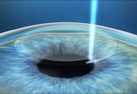
In a single step the VisuMax femtosecond laser creates a thin lenticule and a small access measuring less than 4 mm in the intact cornea.
-

Step 2:
The surgeon removes the lenticule through the small access. There is minimal disruption to the biomechanics of the cornea. No flap needs to be cut.
-

Step 3:
The minimally invasive removal of the lenticule changes the shape of the cornea, correcting the refractive error of the eye.
Laser surgery techniques for refractive vision correction were well established, both scientifically and clinically, by the mid-80s. The basic principle behind all the methods is using a laser to model the outer corneal layer so that the focal point of the bundled incidental light is on the surface of the retina itself and not in front of or behind it.
The original treatment methods, PRK and LASEK, manually removed the outer protective layer (epithelium) of the cornea before the laser surgery.
The recovery process often included postoperative discomfort and very slow improvement of visual acuity.
As a consequence, laser surgery techniques which involved preparing a corneal flap (LASIK and FemtoLASIK) were then developed.
Here, a mechanical cutting tool (microkeratome) or a femtosecond laser is used to create a thin flap of cornea which is just 120 micrometers thick. Almost a complete circle (270°) is cut in the upper layers of the cornea. The flap is then folded back like the cover of a book. Subsequently, the exposed corneal tissue is then ablated using an excimer laser in a separate stage of treatment. The greater the level of vision defect, the more tissue needs to be removed. In the final stage of treatment, the corneal flap is then folded back into its original position. It then adheres in the original place over the coming weeks and months. The disadvantage is that the flap can shift or even tear off (flap displacement) as the result of rubbing or other physical contact after the operation.
The ReLEx® SMILE principle:
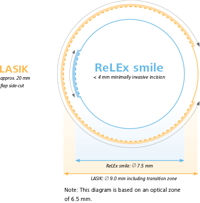
The high-precision VisuMax® femtosecond laser creates a small lens (lenticule) inside the intact cornea, the volume and form of which are determined by the degree of vision defect to be corrected. This lenticule is then removed in a minimally invasive procedure from the interior of the cornea through a small access measuring just a few millimetres. In contrast to earlier techniques, it is no longer necessary to fold back the cornea. There is no flap incision, the area of the incision is reduced to a minimum and the outer corneal layers remain more or less intact. All of which is made possible by a sensitive, precise and convenient treatment method.
Free yourself from glasses and contact lenses!
Register today!
The benefits of ReLEx SMILE
The moment flapless surgery becomes clearly visible: in a smile. This is the moment we work for.
In recent years minimally invasive diagnostic and treatment methods have set new standards in modern medicine. This is because minimally invasive very often means minimum discomfort for the patient. In its ReLEx® SMILE method Carl Zeiss has developed a new technique for the minimally invasive laser correction of vision defects. This innovative treatment technique yields a whole range of benefits:
Flapless treatment
- No folding back of a corneal flap Lenticule preparation in the intact cornea
- Minimally invasive access measuring just a few millimeters (80 % less lateral incision than in LASIK and FemtoLASIK)
- Almost complete preservation of protective corneal layer (epithelium) and stabilizing outer corneal layers (Bowman’s membrane)
- Almost complete preservation of protective corneal layer (epithelium) and stabilizing outer corneal layers (Bowman’s membrane)
- Practically painless during and after the procedure
Entire treatment carried out using femtosecond laser (all-femto)
- Use of high-precision femtosecond technology
- Outstanding predictability of results, even in cases of severe myopia (> -7.00 D)
- Anatomically shaped contact glass avoids unnecessary compression of the cornea
- No loss of vision during treatment
- Entire laser correction in a single treatment step
- No device change during the operation
- Short procedure
- Noiseless and odorless treatment stabilization of visual acuity typically within 14 days

Testimonials

Käroli Michelson
The next morning the world was clear, my eyes didn't hurt and the eyesight was clear.

Liisi Värv
Laser eye surgery was one of the best investments I've made! The people there were friendly sweet and kind. The service was super!

Kaspars U.
After almost 20 years I see without glasses again. Now, from time to time, I imagine why I didn't do it faster.
In and out of focus –
Types of vision defect
The physical/optical principles behind the human eye are similar to those in a camera. The cornea and lens assume the role of the camera lens. They bundle the parallel incidental light rays and determine the focal distance. In an eye with normal vision, the light rays are focused so that the focal point is on the retina itself. The result is a sharply focused image. This is then transmitted via the optic nerve to the brain.
Nearsightedness is the most common vision defect world-wide. Almost half the global population is – to varying degrees – affected by it. In nearsighted people, the eye is too long in relation to its refractive power. Light rays are refracted by the cornea and the lens in such a way that the focal point lies in front of the retina. By the time the rays hit the retina itself, they are already drifting apart. The result is a retinal image which is out of focus. Distant objects appear blurred. Depending on the degree of vision defect, near objects are in sharp focus.
In farsighted people, the eye is too short in relation to the refractive power. Light rays are refracted by the cornea and the lens in such a way that the focal point is behind the retina. A blurred image is then created on the retina because the rays are not yet focused when they hit it. Up to a certain age this lack of refractive power can be compensated by changing the shape of the lens (accommodation). Depending on the extent of the farsightedness, objects which are close, and even distant ones in some cases, are no longer in sharp focus.
In people with astigmatism, the curvature of the cornea is uneven. The resulting refraction causes multiple focal points to be created. Objects both near and far appear skewed or distorted. Astigmatism can occur independently or be accompanied by farsightedness or nearsightedness.

Other things you might like to know about ReLEx SMILE
Free yourself from glasses and contact lenses!
Register today!


